
The J@blation System
The J@blation System represents a new, targeted approach to the treatment of facet joint-related spine disorders. Specifically engineered to treat the posterior synovial capsule and nerve receptors of the facet joints, J@blation utilizes high-frequency radiofrequency (HF/RF) energy to deliver precise thermal therapy. This system is designed to ablate nerve receptors while preserving surrounding structures, offering a focused and minimally invasive solution for facet joint syndrome.
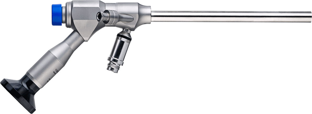
With direct visualization made possible by the MaxMoreSpine Tom Endo Stick Endoscope, the J@blation system ensures a high level of accuracy and safety throughout the procedure. Its three-effect therapy, adjustable based on patient BMI, allows for personalized treatment, while the automated control of therapy time adds consistency and ease of use for the physician.
J@blation stands at the forefront of minimally invasive spine treatment innovation. Offering a simplified, repeatable, and highly effective method to manage facet joint pain, especially in patients where traditional treatments may be less suitable. This makes it an attractive alternative for both patients and clinicians seeking precision, safety, and efficiency in spinal care.
The J@blation procedure
The physician designed and tested J@blation system incorporates procedural simplicity, including:
- Minimally invasive procedure
- Three Effects, based on patients BMI, designed to deliver precise therapy for each patient
- Automatic control of therapy time
- Innovative treatment for facet joint syndrome
The J@blation system is exclusively designed as a system to treat facet joints.
- Place the patient in a prone position, drape and prepare the treatment site(s) following standard techniques.
- Determine desired treatment location via fluoroscopy, and mark skin.
- Make a 10 – 15mm incision in a location that takes into account the angle of approach required to reach the targeted facet joint.
- Using fluoroscopic guidance, advance the K-wire into the incision until it reaches the facet joint. Tap the wire gently into the bone using a small hammer. Confirm via fluoroscopy the correct position of the K-wire.
- Insert the Dilator over the K-wire and confirm via fluoroscopy the correct position of K-wire and Dilator.
- Insert the Working Sleeve over the Dilator / K-wire
- Remove the Dilator / K-wire and insert the Endoscope in the Working Channel to get an overview on the capsular / joint
- Remove the Endoscope and insert the drill to shave the capsular / joint
- Remove the drill and insert the Endoscope and RF Probe and coagulate the capsular
- Remove all instruments and close the incision using standard techniques.
- Move to the next treatment location and repeat steps 1 – 10.
Content Starter Kit
| ARTICLE CODE | PRODUCT DESCRIPTION | QTY |
|---|---|---|
| 1002-TS 004 | Tom Endo Stick | 1 |
| 1001-BB 001 | MaxMoreSpine ® Drill | 1 |
| 1001-ES 25 | Working Sleeve, length112mm / O.D 9.5mm / I.D 8.5mm / O° cut with docking teeth | 1 |
| 1001-GW 006 | Guide Wire, I.D 1.5mm, length 255mm, sharp point on one side | 1 |
| 1001-DC 013 | Dilator, length 170mm, cannulated 1.6mm, O.D 8.4mm | 1 |
| 1001-BH 001 | Ball Handle | 1 |
| 1001 SF-001 | Grasping Forceps, 3.0mm | 1 |
| 1001 ES-19 | Working Sleeve I.D. 8.5mm | 1 |
Download our complete product catalogue here

MaxMore for Physicians
If you’re interested in learning endoscopic techniques or seeking more precise endoscopic instrumentation, use the following links to find out how our solutions fit your needs.

MaxMore for Patients
If you’re a candidate for spinal surgery, use the following links to learn more about endoscopic spine surgery and how to find a local physician performing the maxmorespine® procedure.